In the movie Gattaca, its a space agency that is supposed to be in the near future. At this agency, they are planning a trip to Saturn's moon Titan. In order to take the journey, you must have the right genetic makeup. Vincent Freeman wants badly to make the trip, but in Gattaca he is considered an "in-valid", which means he has defective genes. Vincent "switches" places with Jerome Morrow, right down to the nucleic acid. Vincent takes hair, blood, and urine samples from the real Jerome, to pass all of the required tests at Gattaca. Due to the fact that the real Jerome Morrow was crippled in some kind of accident, Jerome needs Vincent just as much as Vincent needs Jerome.
The scientific premise, like in most science-fiction movies, combines a mix of truth and fiction. In the movie, the alleles from parents are so chosen that the combination produces the optimal arrangement in terms of the child's genotype. But we know enough right now to realise that even in situations where there is a great degree of genetic predisposition, it is quite probable that that predisposition (positive or negative) is never realised. For more complex behavioural traits such as intelligence, aptitude test results would be a far better indicator than genetic makeup. In other words, any correlation people may find between a complex behavioural trait such as intelligence and genetics is for all practical purposes controlled by the environment, given the "edge of chaos" nature of such traits.
"What is disturbing about our genetic engineering capabilities today is no more disturbing than our medical engineering capabilities (and there are plenty of disturbing ramifications). The genetic component simply provides one additional way to discriminate in the real world, just as it is routinely done with age, sex, years of experience, education, and physical ability: Consider the physical and mental requirements for being an astronaut today, or even for admission to college. Information, of any sort, is a valuable commodity in this day and age, and the kind encoded in DNA is no exception. Humans naturally use information to discriminate. I would argue that for some of scenarios posited in Gattaca, the genetic information is far less reliable than physiological and psychological histories. "
Information from:
http://www.ram.org/ramblings/movies/gattaca.html
Monday, April 11, 2011

Friday, April 1, 2011
Bartonella Henselae
While doing a virtual lab on bacterial DNA, I was able to be a lab technician in a molecular biology lab and was responsible for providing lab results for use in diagnosing patients. On the certain DNa that I found, it came up with the bacteria Bartonella Henselae. Here is what was found:
Bartonella henselae Various species of Bartonella that are pathogenic to humans are transmitted via a vector, or directly from an animal reservoir. For example, B. bacilliformis via sandflies causes Oroya fever; B. quintana via body lice causes trench fever; and B. henselae via cats causes cat scratch disease (CSD). CSD typically manifests as swellings of the lymph glands, possibly with skin lesions at the site of inoculation and possibly accompanied by fever, fatigue, and other symptoms. Immunocompromised patients may be particularly susceptible and can develop a different disease, bacillary angiomatosis, as a result of infection by B. henselae or B. quintana.
Below are pictures of Bartonella Henselae:


Bartonella henselae Various species of Bartonella that are pathogenic to humans are transmitted via a vector, or directly from an animal reservoir. For example, B. bacilliformis via sandflies causes Oroya fever; B. quintana via body lice causes trench fever; and B. henselae via cats causes cat scratch disease (CSD). CSD typically manifests as swellings of the lymph glands, possibly with skin lesions at the site of inoculation and possibly accompanied by fever, fatigue, and other symptoms. Immunocompromised patients may be particularly susceptible and can develop a different disease, bacillary angiomatosis, as a result of infection by B. henselae or B. quintana.
Below are pictures of Bartonella Henselae:

How I was able to sequence the DNA:
In the first step, my objective was to:
- Prepare a sample from a patient and isolate whole bacterial DNA.
- Make many copies of the desired piece of DNA.
- Sequence the DNA.
- Analyze the sequence and identify the bacteria.
Saturday, March 19, 2011
In Sickness and In Health
While doing the in sickness and health activity, it was found interesting that just because some people in your family might have a certain disease, there isnt a set chance that you or anyone else may get it. It was neat to see that some of them were just a spark and may not have appeared for any certain reason. While others either went to each generation or maybe even skipped a few but still continued a selevt pattern. It was amazing to see how different they all work.
Review:
While studying the information of the sickness and health, if a person was to have below 1% of the normal level of VIII, the smallest of cuts can cause uncontrolled internal bleeding. Why is this? Due to the fact of having a low level of VIII, causes the blood to be super thin. There is an injection of factor VIII that helps the blood to clot, making it less likely to cause uncontrolled bleeding. Internal bleeding, can cause complications such as: swelling, joint damage, and even an increased chance of neurological complications. Very simple procedures can become too risky for a person with a low level of VIII. A person can either receive the injection of factor VIII monthly, or twice a year at a clinic. This is a therapy called prophylaxis.
A little bit more about factor VIII, its in the hemostatic system where the blood vessels have a huge role in a human beings survival. Hemophilia is the linked pattern of inheritance of bleeding diathesis. Due to the deficiency of factor F in the blood, hemophilia is caused. There was a test in the early 1900's and results found that normal plasma could shorten the blood clotting time of the hemophilic blood. It was soon found that a factor present in normal plasma showed to be effective to accelerate coagulation of the hemophilic blood. This was soon names the Factor VIII-C. Further testing is still to this day being tested and explored to see the roles of increased levels of factor VIII in a thrombophilic state. Factor VIII is a largely inherited disorder, with the proteins that are needed to form blood clots in the body is either missing or reduced. Studies show that around 30% of the cases of factor VIII show no family history of this disorder, although its the result of a spontaneous gene mutation. In some cases, the gene cane be "hidden". Numbers show around 1 in every 5,000 males have the factor. It doesnt affect a certain race or gender. The hemophilia gene is located on the X chromosome which everyone inherits. There is a product for people with VIII that is called desmopressin acetate (DDAVP) in order to treat small vuts.
Found this cool little bit of information!
Normal plasma levels of FVIII range from 50% to 150%. There are different levels of hemophilia: mild, moderate, and severe, depending on the amount of clotting factor in the blood:
People with mild hemophilia have 5% up to 50% of the normal clotting factor in their blood. Most patients usually have problems with bleeding only after serious injury, trauma or surgery. In many cases, mild hemophilia is not diagnosed until an injury, surgery or tooth extraction results in prolonged bleeding. The first episode may not occur until adulthood. Women with mild hemophilia often experience menorrhagia, heavy menstrual periods, and can hemorrhage after childbirth.
People with moderate hemophilia about, 15% of the hemophilia population, have 1% up to 5% of the normal clotting factor in their blood. They tend to have bleeding episodes after injuries and some without obvious cause. These are called spontaneous bleeding episodes.
People with severe hemophilia about 60% of the hemophilia population, have <1% of the normal clotting factor in their blood. They have bleeding following an injury and may have frequent spontaneous bleeding episodes, often into their joints and muscles
There are four possible outcomes for the baby of a woman who is a carrier. These four possibilities are repeated for each and every pregnancy:
1. A girl who is not a carrier
2. A girl who is a carrier
3. A boy without hemophilia
4. A boy with hemophilia
Review:
While studying the information of the sickness and health, if a person was to have below 1% of the normal level of VIII, the smallest of cuts can cause uncontrolled internal bleeding. Why is this? Due to the fact of having a low level of VIII, causes the blood to be super thin. There is an injection of factor VIII that helps the blood to clot, making it less likely to cause uncontrolled bleeding. Internal bleeding, can cause complications such as: swelling, joint damage, and even an increased chance of neurological complications. Very simple procedures can become too risky for a person with a low level of VIII. A person can either receive the injection of factor VIII monthly, or twice a year at a clinic. This is a therapy called prophylaxis.
A little bit more about factor VIII, its in the hemostatic system where the blood vessels have a huge role in a human beings survival. Hemophilia is the linked pattern of inheritance of bleeding diathesis. Due to the deficiency of factor F in the blood, hemophilia is caused. There was a test in the early 1900's and results found that normal plasma could shorten the blood clotting time of the hemophilic blood. It was soon found that a factor present in normal plasma showed to be effective to accelerate coagulation of the hemophilic blood. This was soon names the Factor VIII-C. Further testing is still to this day being tested and explored to see the roles of increased levels of factor VIII in a thrombophilic state. Factor VIII is a largely inherited disorder, with the proteins that are needed to form blood clots in the body is either missing or reduced. Studies show that around 30% of the cases of factor VIII show no family history of this disorder, although its the result of a spontaneous gene mutation. In some cases, the gene cane be "hidden". Numbers show around 1 in every 5,000 males have the factor. It doesnt affect a certain race or gender. The hemophilia gene is located on the X chromosome which everyone inherits. There is a product for people with VIII that is called desmopressin acetate (DDAVP) in order to treat small vuts.
Found this cool little bit of information!
Normal plasma levels of FVIII range from 50% to 150%. There are different levels of hemophilia: mild, moderate, and severe, depending on the amount of clotting factor in the blood:
People with mild hemophilia have 5% up to 50% of the normal clotting factor in their blood. Most patients usually have problems with bleeding only after serious injury, trauma or surgery. In many cases, mild hemophilia is not diagnosed until an injury, surgery or tooth extraction results in prolonged bleeding. The first episode may not occur until adulthood. Women with mild hemophilia often experience menorrhagia, heavy menstrual periods, and can hemorrhage after childbirth.
People with moderate hemophilia about, 15% of the hemophilia population, have 1% up to 5% of the normal clotting factor in their blood. They tend to have bleeding episodes after injuries and some without obvious cause. These are called spontaneous bleeding episodes.
People with severe hemophilia about 60% of the hemophilia population, have <1% of the normal clotting factor in their blood. They have bleeding following an injury and may have frequent spontaneous bleeding episodes, often into their joints and muscles
There are four possible outcomes for the baby of a woman who is a carrier. These four possibilities are repeated for each and every pregnancy:
1. A girl who is not a carrier
2. A girl who is a carrier
3. A boy without hemophilia
4. A boy with hemophilia
Monday, March 14, 2011
DNA
One may ask, what exactly is DNA? Well, its the hereditary material in humans and almost all other organisms. Almost all cells in the human body have some sort of DNA. Where is it located? DNA is mostly located in the cell nucleus where it is also called nuclear DNA. Although most of DNA is located in the cell nucleus, there can also be some found in the mitochondria. Here it is called mitochondrail DNA. DNA is made up of 4 chemical bases. (A), guanine (G), cytosine (C), and thymine (T). Human DNA is composed of 3 billion bases. Over 99% of those bases are the same in every human being. The order determines the amount of information available. Which is used for building and maintaining an organism. Each DNA bases pairs up with one another. A with T and C with G, to form units that are called base pairs. Every base is attached to a sugar molecule and a phosphate molecule. Altogether, a base, sugar, and phosphate are called a nucleotide. Nucleotides are in strands of two that form a spiral which is called a double helix. The double helix may look something like a ladder. The base pairs form the ladder's rungs, and the sugar and phosphates form the vertical sidepieces. DNA is able to replicate, also known as make copies of itself. Each and every strand of DNA in a double helix serves as a pattern for duplicating in the sequence of bases. This is very critical in cell division, due to the fact that each new cell needs an exact copt of the DNA present in the old cell.


Thursday, January 20, 2011
Meiosis

Meiosis is what is known as a type of cell division in which the germ cells (eggs and sperm) are produced. Meiosis involves a reduction in the number of genetic materials. Two successive nuclear divisions with only one round of DNA replication is comprised in meiosis.
Interphase: Before meiosis begins, genetic material is duplicated.
First division of meiosis
Prophase 1: Duplicated chromatin condenses. Each chromosome consists of two, closely associated sister chromatids. Crossing-over can occur during the latter part of this stage.
Metaphase 1: Homologous chromosomes align at the equatorial plate.
Anaphase 1: Homologous pairs separate with sister chromatids remaining together.
Telophase 1: Two daughter cells are formed with each daughter containing only one chromosome of the homologous pair.
Second division of meiosis: Gamete formation
Prophase 2: DNA does not replicate.
Metaphase 2: Chromosomes align at the equatorial plate.
Anaphase 2: Centromeres divide and sister chromatids migrate separately to each pole.
Telophase 2: Cell division is complete. Four haploid daughter cells are obtained.
Four daughter cells are produced by one parent cell. Daughter cells have halod of the number of chromosomes found in the original parent cell , and due to crossing over are genetically different.
info from: http://www.accessexcellence.org/ and class discussions
Self Reflection
During the third quarter this year, there are a lot of things that I wish to learn. I'd like to get more information on mitosis, stem cells, meiosis, and so one. While learning this things, I hope that they will help my throughout the rest of my life, as I hope to be able to go in any direction from high school. I believe that learning this things and many more will benefit me in the long run. I will have a lot more outside knowledge!
Monday, January 17, 2011
Mitosis!!! :) Finished and Improved!!
There are 5 different stages of mitosis;
1.) interphase
2.) prophase
3.) metaphase
4.) anaphase
5.) telophase
While doing the onion tip activity, I found the following information:


1.) interphase
2.) prophase
3.) metaphase
4.) anaphase
5.) telophase
While doing the onion tip activity, I found the following information:
|

Mitosis In Onion Root Tips

A mechanism is mitosis that allows the nuclei of cells to split and in return provide each of teh daughter cells with a full set of chromosomes during cellular division. This along with cytokinesis occur in all multicellular plants and animans to permit the growth of the organism, Cytokinesis is the division of cytoplasm.
Resting Cell
This normal resting cell is in a state called interphase, which the chromatin is undifferentiated in the heavily-stainded nucleus. (shown above). In order for a cell to enter the mitosis phase, it must first undergo a synthesis phase, which is where each chromosome is duplicated and consists of two sister chromatids that are joined together. Prophase is the first phase of mitosis. This is where the nuclear chromatin starts to become more organized and then condenses into a thick strand that will eventually become a chromosome.
Early Prophase
Late prohase, or what is known as prometaphase usually begins with the disruption of the nuclear envelope. This is broke down into small membrane vesicles that are very similar to the endoplasmic reticulum, and can tend to remain visible around the mitotic spindle. During this time, the chromosomes continue to condense and then gradually shorten and thicken until they are completely formed units that will soon undergo mitosis. The nucleolus will also disappear during this stage.
Late Prophase
Metaphase is where the chromosomes that are attached to the kinetochore microtubles begin to align with one plane known as the metaphase plate, which are located half way between the spindle poles. Due to the tension exerted by the kinetochore microtubules on the chromosomes and the entire spindle cromosome complex, we are now ready for the next event. The photo below shows a onion root tip cell chromosomes in metaphase, ready for seperation. You can clearly see the kinetochore and polar microtubules and they radiate out the ends of the cell, leaving the chromosomes in the middle.

Metaphase
Next around, the chromosomes are set up for seperation into the next stage called anaphase. Shortly after the metaphase chromosomes are alighned at the metaphase plate, two halves of each chromosome are pulled apart by the spindle apparatus and migrate to the opposite spindle poles. As the kinetochore microtubules are shortened, the chromosomes are pulled toward the poles with the polar microtubules elongate to assist in the seperation process.
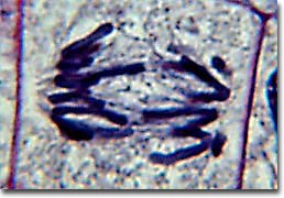
Early Anaphase
Anaphase is known as a rapid process that lasts only a few short minutes. When chromosomes have completely migrated to the spindle poles, the kinetochore microtubules start to disappear while the polar microtubules continue to elongate. This is known as the junction between late anaphase and early telophase, which is the final step in chromosome division.

Late Anaphase
Telophase, the daughter chromosomes arrive at the spindle poles and are eventually redistributed into chromatin. Cytokinesis is the process in which the cytoplasm is divided by cleavage, and also starts sometime in late anaphase and continues through telophase. When the chromosomes and their extrusion to the spindle poles are completely separated, the nuclear membrane starts to reform around each group of chromosomes at the opposite ends of the cell. The nucleoli will also start to reappear in what will soon be the two new cell nuclei.

Telophase
After telophase is complete, the new cell membrane is now being formed. The nuclei have almost completely matured to the pre-mitotic state. The last steps include the completion of the total formation of a membrane between each of the new daughter cells to yield them to two separate new cells.

Metaphase
Next around, the chromosomes are set up for seperation into the next stage called anaphase. Shortly after the metaphase chromosomes are alighned at the metaphase plate, two halves of each chromosome are pulled apart by the spindle apparatus and migrate to the opposite spindle poles. As the kinetochore microtubules are shortened, the chromosomes are pulled toward the poles with the polar microtubules elongate to assist in the seperation process.
Early Anaphase
Anaphase is known as a rapid process that lasts only a few short minutes. When chromosomes have completely migrated to the spindle poles, the kinetochore microtubules start to disappear while the polar microtubules continue to elongate. This is known as the junction between late anaphase and early telophase, which is the final step in chromosome division.
Late Anaphase
Telophase, the daughter chromosomes arrive at the spindle poles and are eventually redistributed into chromatin. Cytokinesis is the process in which the cytoplasm is divided by cleavage, and also starts sometime in late anaphase and continues through telophase. When the chromosomes and their extrusion to the spindle poles are completely separated, the nuclear membrane starts to reform around each group of chromosomes at the opposite ends of the cell. The nucleoli will also start to reappear in what will soon be the two new cell nuclei.
Telophase
After telophase is complete, the new cell membrane is now being formed. The nuclei have almost completely matured to the pre-mitotic state. The last steps include the completion of the total formation of a membrane between each of the new daughter cells to yield them to two separate new cells.
Daughter Cells
Subscribe to:
Posts (Atom)